Genetic Testing Methods:
Testing Options
After the egg has been fertilized, PGD can be performed. PGD involves the removal of cellular material and the subsequent testing of the DNA in that material. As PGD techniques have advanced, clinicians can conduct the genetic testing at various time points post-fertilization. Depending on when genetic analysis is conducted, the number of cells removed may change. Today PGD can be conducted at one of three stages of embryonic development and clinics vary with regard to the timing and type of genetic testing. (image 2)
PGD can be performed at:
Day 1 Polar Bodies – by the removal of a polar body, which is a byproduct of the first cell division post fertilization.

Day 3 Blastomere (8 cells) – by the removal of 1-2 cells of the early embryo.

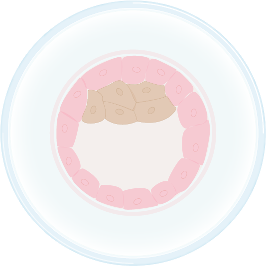
Day 5 Blastocyst (150 cells) – by the removal of multiple cells from the the outer layer of the embryo called the trophoblast, which will go on to become the cells of the placenta, while the inner cells go on to become the embryo.
Types of genetic visualization for PGD
Since the amount of genetic material that is taken from the embryo is a very small quantity, visualization techniques have been developed that allow detailed genetic analysis. Polymerase Chain Reaction (PCR) is a laboratory technique that amplifies a small amount of DNA from a single cell or small number of cells and allows for genetic testing outside of the cells. For dominant mutations like BRCA1/2, it is important to test two cells from the embryo using PCR. Another type of genetic visualization is Fluorescent in-situ hybridization (FISH), a technique commonly used to detect chromosomal abnormalities (eg. Trisomy 21). FISH involves the binding of specific DNA probes to regions of chromosomes in the cells.